Deltoid Anatomy
Updated:
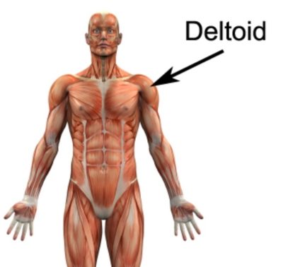
The deltoid muscle is a significant component of the human shoulder complex. Comprised of three distinct portions, the anterior (front), middle, and posterior (rear) deltoids, this muscle plays a vital role in facilitating a wide range of arm movements. In this article, we will delve into the comprehensive details of deltoid anatomy, including its origin, insertion, action, nerve supply, blood supply, and common injuries. So, let’s explore the intricacies of this remarkable muscle.
Origin and Insertion
The deltoid muscle originates from three distinct areas. The anterior deltoid originates from the lateral third of the clavicle. The middle deltoid arises from the acromion process of the scapula. Lastly, the posterior deltoid originates from the spine of the scapula. These three portions converge to form a common tendon, which inserts onto the deltoid tuberosity of the humerus.
Action
The deltoid muscle is responsible for several essential movements of the arm. The anterior deltoid aids in shoulder flexion, horizontal flexion and medial rotation. The middle deltoid assists in shoulder abduction. Lastly, the posterior deltoid contributes to shoulder extension, horizontal extension and lateral rotation.
Nerve Supply
The deltoid muscle receives its nerve supply from the axillary nerve, which originates from the brachial plexus. This nerve also innervates the teres minor muscle, providing essential motor function to the shoulder joint.
Blood Supply
The deltoid muscle receives its blood supply from branches of the posterior circumflex humeral artery and the deltoid branch of the thoracoacromial artery. These arteries ensure an adequate oxygen and nutrient supply to the muscle fibers, facilitating their proper function.
Function
The deltoid muscle is not only responsible for generating movement in the shoulder joint but also contributes to overall shoulder stability. By contracting and relaxing, the deltoid assists in various activities such as lifting objects, throwing, pushing, and performing overhead movements. Its ability to generate force and control movement makes it crucial for functional activities in daily life and sports performance.
Deltoid Anatomy Palpation
Palpating the deltoid muscle can provide valuable information regarding its size, tone, and any potential abnormalities. To palpate the deltoid, one can start by locating the bony landmarks of the shoulder, including the acromion process and the deltoid tuberosity. By gently pressing and moving the fingers along the muscle belly, clinicians can assess the muscle’s overall condition, identify trigger points, and detect any signs of tenderness or swelling, aiding in the diagnosis of deltoid-related conditions.
Deltoid Anatomy – Clinical Relevance
Understanding the intricacies of deltoid anatomy has significant clinical relevance in various fields. Healthcare professionals, such as orthopedic surgeons, physical therapists, and sports medicine specialists, rely on this knowledge to diagnose and treat shoulder injuries and conditions. They can employ specific rehabilitation exercises and therapeutic techniques to address deltoid-related issues, such as strains, tears, impingements, and tendinitis. Furthermore, a thorough understanding of deltoid anatomy is essential for proper administration of injections, as certain medication administrations, like deltoid intramuscular injections, require precise localization of the muscle.
By understanding the function, palpation techniques, and clinical relevance of the deltoid muscle, healthcare practitioners can effectively evaluate, treat, and manage conditions affecting the shoulder complex. Moreover, this knowledge empowers individuals to engage in preventive measures and exercise programs that promote deltoid strength and shoulder health, thereby reducing the risk of injuries and improving overall musculoskeletal well-being.
Common Injuries
While the deltoid muscle is generally robust, it can be prone to certain injuries. Some of the most common deltoid injuries include:
- Deltoid Strain: This occurs due to overuse or sudden excessive force on the muscle, leading to micro-tears in the muscle fibers.
- Deltoid Tendinopathy: Inflammation of the deltoid tendon, often caused by repetitive movements or excessive stress on the muscle.
- Deltoid Impingement: Compression of the deltoid muscle and associated structures, usually due to structural abnormalities or poor biomechanics.
- Deltoid Tear: A partial or complete tear of the deltoid muscle, typically resulting from a traumatic event or forceful impact.
- Subdeltoid or Subacromial Bursitis: Although not affecting the deltoid directly, this can be a source of pain arising from inflammation of one or more bursa located underneath the deltoid muscle. Typically occurring due to excessive load on the bursa associated with overuse or trauma.
Deltoid Anatomy – References
- Moore KL, Dalley AF, Agur AMR. Clinically Oriented Anatomy. 8th ed. Philadelphia, PA: Wolters Kluwer; 2017.
- Standring S. Gray’s Anatomy: The Anatomical Basis of Clinical Practice. 41st ed. New York, NY: Elsevier; 2016.
- Deltoid Muscle by KenHub
Conclusion
The deltoid muscle is a remarkable structure that plays a crucial role in arm movement and stability. Understanding its origin, insertion, action, nerve supply, blood supply, and common injuries is essential for healthcare professionals, athletes, and individuals interested in shoulder anatomy. By grasping the intricacies of deltoid anatomy, we can better appreciate the remarkable capabilities and potential vulnerabilities of this crucial shoulder muscle.

Link to this Page
If you would like to link to this article on your website, simply copy the code below and add it to your page:
<a href="https://physioadvisor.com.au/deltoid-anatomy”>Deltoid Anatomy – PhysioAdvisor.com</a><br/>Learn about Deltoid anatomy including origin, insertion, action, nerve supply, bloody supply, common injuries and more on PhysioAdvisor.
Return to the top of Deltoid Anatomy.