Patellar Dislocation
Updated:
(Also known as Patella Dislocation, Dislocated Patella, Knee Cap Dislocation, Knee Dislocation)
What is patellar dislocation?
Patellar dislocation is a relatively common traumatic sporting injury characterized by tearing of the connective tissue surrounding the knee cap (patella) with subsequent displacement of the patella so it is completely out of its normal position.
The knee comprises of the union of 3 bones – the long bone of the thigh (femur), the shin bone (tibia) and the knee cap (patella) (figure 1). The patella (knee cap) is situated at the front of the knee and lies within a groove at the front of the thigh bone. The tendon of the quadriceps muscle (the muscle at the front of the thigh) envelops the patella and attaches to the top end of the tibia (figure 1). Due to this relationship, the knee cap sits in front of the femur forming a joint in which the bones are almost in contact with each other. The surface of each bone, however, is lined with cartilage to allow cushioning between the bones. The patella also has strong bands of connective tissue known as the patella retinaculum (figure 2) attaching the knee cap on either side of the femur. This joint is called the patellofemoral joint.
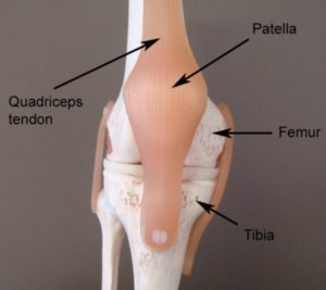
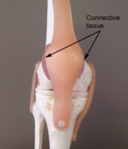
Normally, the patella is aligned in the middle of the patellofemoral joint and is held firmly in place by the quadriceps muscle and patella retinaculum. Occasionally, the patella may be pushed completely out of its normal position. This normally occurs due to traumatic forces pushing the knee cap out of position beyond what the quadriceps and patella retinaculum can withstand. When this occurs the condition is known as patellar dislocation.
Patellar dislocation usually occurs in a direction towards the outside of the knee (i.e. away from the other leg). During dislocation, tearing and disruption of the patellar retinaculum usually occurs. The joint surfaces may also be damaged and occasionally there may be an associated fracture. In many cases of patellar dislocation, the patella spontaneously moves back into its original position often with knee extension (i.e. straightening of the knee).
Causes of patellar dislocation
Patellar dislocation typically occurs when the forces pushing the knee cap out of its normal position are greater than the quadriceps muscle and patella retinaculum can resist. This typically occurs traumatically due to excessive twisting or jumping forces or due to a direct blow (usually to the inner aspect of the patella). Occasionally, however, it may occur in the absence of trauma especially in young girls who are hyper-flexible. Patients with this condition are frequently seen in contact sports or sports requiring rapid changes in direction, such as football or rugby.
Signs and symptoms of patellar dislocation
Patients with this condition usually experience sudden, intense pain at the front of the knee during injury. Pain is usually associated with a feeling of the knee ‘giving way’ or of something ‘popping out’. There may be a noticeable visible deformity of the knee owing to the knee cap moving out of position (normally to the outer aspect of the knee) when compared to the unaffected knee. There may also be a rapid onset of knee swelling within the first 1-2 hours following injury. In patients who have experienced recurrent episodes of patellar dislocation, the knee cap may easily move back into its original position with certain knee movements (normally straightening of the knee). In these cases, pain and swelling may also be relatively minimal.
Once the patella has returned to its original position following dislocation, patients usually experience an ache that may increase to a sharper pain with activity. Pain is typically experienced during activities that bend or straighten the knee particularly whilst weight bearing. Activities that frequently aggravate symptoms include going up and down stairs or hills, squatting, lunging, running, jumping or attempting to kneel. Pain typically increases on firmly touching the borders of the patellofemoral joint (i.e. along the patellar retinaculum – figure 2) There may also be an associated clicking or grinding sound when bending or straightening the knee. Patients may also experience episodes of the knee giving way or collapsing due to pain. In long standing cases, there may be evidence of quadriceps muscle wasting.
Diagnosis of patellar dislocation
A thorough subjective and objective examination from a physiotherapist is usually sufficient to diagnose patellar dislocation. Investigations such as X-ray or MRI should usually be performed to assist with diagnosis and rule out other conditions, such as fractures or severe cartilage damage.
Treatment for patellar dislocation

Members Only ContentBecome a PhysioAdvisor Member to gain full access to this exclusive content. For more details see Become a Member. Already a member? Login Now
Surgery for patellar dislocation
Despite appropriate physiotherapy management, some patients undergoing conservative treatment fail to improve or experience recurrent episodes of dislocation and subsequently require surgery for an optimal outcome. The majority of patients with severe cartilage damage or fractures also require surgery. Surgery for patellar dislocation is usually arthroscopic and typically involves removal of any loose bodies or torn cartilage and reconstruction of the torn patella retinaculum. The treating physiotherapist and doctor will refer to a specialist if surgery may be indicated. Physiotherapy and rehabilitation is required following surgery to ensure an optimal outcome and enable a safe return to activity or sport.
Contributing factors to the development of patellar dislocation
There are several factors which can predispose patients to developing this condition. These need to be assessed and, where possible, corrected with direction from a physiotherapist. Some of these factors include:
- muscle weakness (especially the quadriceps (VMO), gluteals or hamstrings)
- tight lateral (outer) structures, such as the lateral patella retinaculum or iliotibial band (ITB)
- “loose” medial (inner) patella retinaculum
- general hypermobility (ligament laxity)
- a shallow femoral groove (i.e. the groove that the knee cap sits within)
- genu valgum (‘knock knees’)
- femoral anteversion (where the thigh bones turn inward)
- patella alta (abnormally high patella in relation to the thigh bone)
- poor lower limb biomechanics (flat feet, increased Q angle)
- muscle tightness (e.g. vastus lateralis, ITB, hip internal rotators, calfs)
- poor balance
- poor pelvic or core stability
- poor landing strategies
- poor coordination
- gender (i.e. greater likelihood in females)
- certain stages of the menstrual cycle (e.g. at mid-cycle (ovulation) when oestrogen peaks and ligament laxity increases)
- fatigue
Physiotherapy for patellar dislocation
Physiotherapy treatment for patellar dislocation is vital to hasten the healing process, reduce the likelihood of recurrence and ensure an optimal outcome. Treatment may comprise:
- soft tissue massage
- electrotherapy
- the use of crutches
- the use of a knee immobilisation brace
- patella taping or bracing to correct patella position
- mobilization
- dry needling
- ice or heat treatment
- progressive exercises to improve flexibility, balance and strength (especially the VMO muscle)
- hydrotherapy
- activity modification advice
- biomechanical correction
- anti-inflammatory advice
- the use of Real-Time Ultrasound to assess and retrain the VMO muscle
- a gradual return to activity program
Other intervention for patellar dislocation
Despite appropriate physiotherapy management, some patients with this condition do not improve. When this occurs the treating physiotherapist or doctor can advise on the best course of management. This may include pharmaceutical intervention, corticosteroid injection or referral to an orthopaedic specialist who will advise on any procedures that may be appropriate to improve the condition. A review with a podiatrist may also be indicated for the prescription of orthotics to correct any foot posture abnormalities.
Exercises for patellar dislocation
The following exercises are commonly prescribed to patients with this condition. You should discuss the suitability of these exercises with your physiotherapist or orthopaedic surgeon prior to beginning them. Generally, they should be performed 3 times daily and only provided they do not cause or increase symptoms.
Your physiotherapist can advise when it is appropriate to begin the initial exercises and eventually progress to the intermediate, advanced and other exercises. As a general rule, addition of exercises should take place provided there is no increase in symptoms.
Initial Exercises
Static Quadriceps Contraction
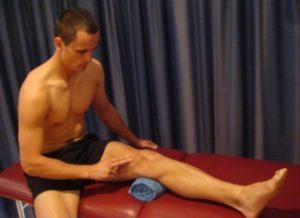
Tighten the muscle at the front of your thigh (quadriceps) by pushing your knee down into a towel (figure 4). Put your fingers on your inner quadriceps to feel the muscle tighten during contraction. Hold for 5 seconds and repeat 10 times as hard as possible without increasing your symptoms.
Adductor Squeeze (Supine)
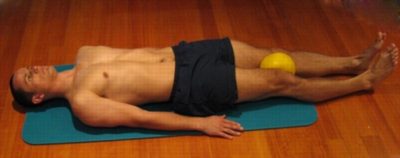
Begin this exercise lying on your back in the position demonstrated with a Pilates ball between your knees (figure 5). Tighten your thigh muscles (quadriceps) by straightening your knees and then slowly squeeze the ball between your knees tightening your inner thigh muscles (adductors). Hold for 5 seconds and repeat 10 times as hard as possible and comfortable provided the exercise is pain free.
Knee Bend to Straighten
Bend and straighten your knee as far as you can go without pain and provided you feel no more than a mild to moderate stretch (figure 6). Gradually increase movement as tolerated provided the exercise is pain free. Repeat 10 – 20 times provided there is no increase in symptoms.
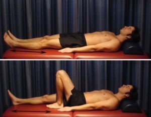
Hip Extension in Standing
Begin this exercise standing at a table or bench for balance. Keeping your back and knee straight, slowly take your leg backwards, tightening your bottom muscles (gluteals) (figure 7). Hold for 2 seconds then slowly return to the starting position. Repeat 10 times provided the exercise is pain free.
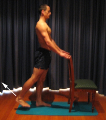
Intermediate Exercises

Members Only ContentBecome a PhysioAdvisor Member to gain full access to this exclusive content. For more details see Become a Member. Already a member? Login Now
Advanced Exercises

Members Only ContentBecome a PhysioAdvisor Member to gain full access to this exclusive content. For more details see Become a Member. Already a member? Login Now
Other Exercises

Members Only ContentBecome a PhysioAdvisor Member to gain full access to this exclusive content. For more details see Become a Member. Already a member? Login Now
Rehabilitation protocol for patella dislocation

Members Only ContentBecome a PhysioAdvisor Member to gain full access to this exclusive content. For more details see Become a Member. Already a member? Login Now
 Find a Physio for patellar dislocation
Find a Physiotherapist in your local area who can treat patellar dislocation.
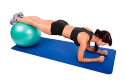 More Exercises
- View more Knee Strengthening Exercises.
- View more Knee Flexibility Exercises.
- View more Balance Exercises.
- View more Leg Stretches.
- View more Leg Strengthening Exercises.
 Physiotherapy products for patellar dislocation
Some of the most commonly recommended products by physiotherapists to hasten healing and speed recovery in patients with patellar dislocation include:
-
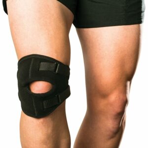 AllCare Ortho Patella Tracker (AOK28)
 -
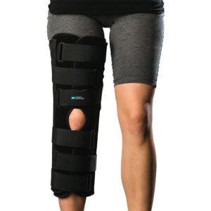 AllCare Ortho – Tri-Panel Knee Immobiliser (AOK81)
 -
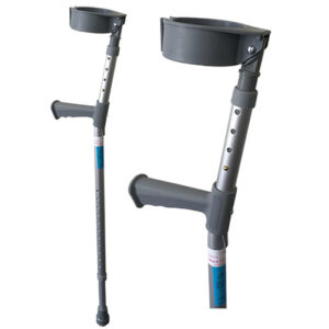 Forearm Crutches Adjustable – Standard Grip
 -
 AllCare Band
 -
 Premium Strapping Tape 38mm (Victor)
 -
 AllCare Tubing
 -
 AllCare Spikey Massage Ball
 -
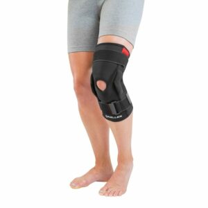 Mueller Hinged Knee Brace
 -
 Fixomull Stretch 5cm x 10m
 -
 AllCare Instant Cold Pack (15 x 25cm)
 -
 AllCare Foam Roller Round
To purchase physiotherapy products for patellar dislocation click on one of the above links or visit the PhysioAdvisor Shop.
 More Information
- View detailed information on initial injury management and the R.I.C.E. Regime.
- View detailed information on How to use Crutches.
- View detailed information on when to use Ice or Heat.
- View detailed information on a Return to Running Program.
- View detailed information on how to safely Return to Sport.
- View detailed information on Patella Taping.
- Unsure of your most likely diagnosis? View our Knee Diagnosis Guide.
Become a PhysioAdvisor Member

Link to this Page
If you would like to link to this article on your website, simply copy the code below and add it to your page:
<a href="https://physioadvisor.com.au/injuries/knee/patellar-dislocation”>Patellar Dislocation – PhysioAdvisor.com</a><br/>Patellar Dislocation - Explore causes, signs and symptoms, diagnosis, treatment, exercises, rehabilitation protocol, physiotherapy products and more.
Return to the top of Patellar Dislocation.

