Diagnostic Guide – Knee Pain
Updated:
Patients suffering from knee pain are commonly seen in clinical practice. Pain may be caused by local structures within or around the knee, or, may be referred from other sources (such as the lower back or hip joint).
Sudden onset knee pain often occurs in athletes involved in fast moving change-of-direction sports, kicking sports, contact sports and those sports which involve twisting movements or rapid acceleration and deceleration (such as football, soccer, basketball, netball and alpine skiing). These acute injuries often involve tearing of cartilage tissue or ligaments within the region. One of the most common causes of sudden onset pain in the knee is a Medial Meniscal Tear (figure 1) often presenting as pain at the inner aspect of the knee.
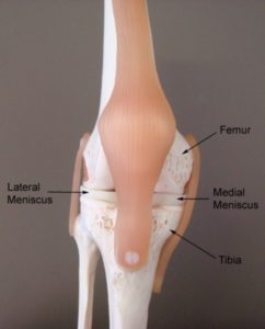
Gradual onset knee pain often occurs in those patients involved in sports or activities that involve high running loads, repetitive squatting, lunging, jumping, twisting, kneeling or repetitive kicking. One of the most common causes of gradual onset pain located at the front of the knee is Patellofemoral Pain Syndrome. Patients with gradual onset pain located at the inner or outer aspect of the knee are often due to either a Medial Meniscal Tear or Lateral Meniscal Tear (Figure 1). In older patients with gradual onset knee pain, the most likely cause of symptoms is degenerative changes in the knee or patellofemoral joint, such as Knee Osteoarthritis.
There are numerous other causes of knee pain, some of which present suddenly due to a specific incident, others which develop gradually over time.
Below are some of the more common causes of knee pain with a brief description of each condition to aid diagnosis. Conditions have been organised according to sudden or gradual onset and common or less common conditions for ease of use.
Find out what may be causing your knee pain:
Sudden Onset Knee Pain – Common Injuries
Medial Meniscal Tear
Damage to cartilage located at the inner aspect of the knee joint (figure 1 – medial meniscus) usually due to excessive weight bearing or twisting forces. Pain is usually located at the inner aspect of the knee and often increases on firmly touching a specific area of the inner knee joint line. Swelling and a clicking, catching or locking sensation may also be present. Symptoms are usually exacerbated with excessive weight-bearing, deep squatting and twisting activities.
Lateral Meniscal Tear
Damage to cartilage located at the outer aspect of the knee joint (figure 1 – lateral meniscus) usually due to excessive weight bearing or twisting forces. Pain is usually located at the outer aspect of the knee and often increases on firmly touching a specific area of the outer knee joint line. Swelling and a clicking, catching or locking sensation may also be present. Symptoms are usually exacerbated with weight-bearing, deep squatting and twisting activities.
MCL Tear
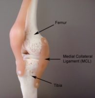
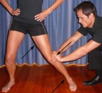
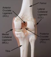
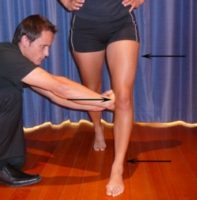
Tearing of the medial collateral ligament of the knee (MCL – Figure 2) typically following a valgus force (Figure 3) or twisting movement and often associated with a snap or tearing sensation at the time of injury. Associated with pain on firmly touching the MCL and often swelling. Occasionally associated with knee instability or giving way of the knee.
ACL Tear
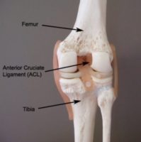
Tearing of the anterior cruciate ligament of the knee (ACL – Figure 4) typically following landing from a jump, sudden deceleration, or due to a hyperextension, sideways or twisting force. Often associated with a ‘snap’, ‘pop’ or ‘tearing’ sensation or feeling of ‘something going out and then going back’ at the time of injury. Usually associated with severe knee pain at the time of injury, significant swelling within a few hours of injury and a feeling of knee instability or giving way of the knee during certain movements.
PCL Tear
Tearing of the posterior cruciate ligament of the knee (PCL – Figure 5) typically following knee hyperextension, or a direct blow to the front of the shin. Often associated with a ‘snap’, ‘pop’ or ‘tearing’ sensation at the time of injury and poorly defined knee pain often most prominent at the back of the knee. Swelling may be minimal and there may be a feeling of knee instability, giving way or a ‘jelly-like’ feeling in the knee during certain movements.
Patellar Dislocation
Movement of the knee cap completely out of its normal position (usually to the outer aspect of the knee) typically following trauma such as a landing from a jump, a twisting movement or a direct blow to the side of the knee cap. May also occur without trauma particularly in young girls who are hyper mobile. Usually associated with severe pain at the front of the knee, deformity, loss of function and often swelling.
Less Common Sudden Onset Injuries
LCL Tear
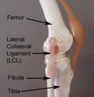
Tearing of the lateral collateral ligament of the knee (LCL – Figure 6) typically following a varus force (Figure 7) or twisting movement and often associated with a snap or tearing sensation at the time of injury. Associated with knee pain located at the outer aspect of the knee, pain on firmly touching the LCL and often swelling. Occasionally associated with knee instability or giving way of the knee.
Acute Patellofemoral Joint Injury
Damage to the knee cap (patella) or cartilage lining the joint between the knee cap and thigh bone (femur) (figure 8) often due to a fall onto the knee cap or direct blow to the front of the knee. Pain is usually felt at the front of the knee and may increase during weight-bearing, squatting or kneeling activities.
Referred Pain
Pain referred to the knee from another source such as the lower back or hip joint, often associated with symptoms above or below the knee (such as lower back pain or stiffness or pain in the hip, buttock, groin, thigh, lower leg, ankle or foot). Typically associated with pain on firmly touching the region responsible for the referred pain or loss of movement in that region. Sometimes in association with pins and needles or numbness in the affected leg or foot.
Acute Fat Pad Impingement
Compression of fatty tissue located at the front of the knee directly below the knee cap, usually due to a knee hyperextension injury. Typically associated with significant knee pain, pain on firmly touching the region directly below the knee cap and exacerbation of symptoms during knee extension activities.
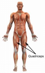
Low Quadriceps Contusion
Bruising of the lower aspect of the front of the thigh (quadriceps – figure 9) following a direct impact from an object or person. Typically associated with pain on firmly touching the affected region.
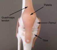
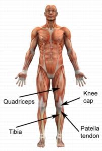
Patellar Tendon Rupture
Rupture of the patellar tendon (figure 10) associated with sudden onset pain in the front of the knee often with an associated ‘snap’ or ‘tearing’ sensation at the time of injury. Usually due to a specific incident such as a landing from a jump or when stumbling. Associated with marked loss of function such as an inability to stand or straighten the knee. Swelling and loss of fullness at the front of the knee is typically present.
Superior Tibiofibular Joint Dislocation
Separation of the joint where the outer lower leg bone (fibula) joins the inner lower leg bone (tibia) just below the knee joint at the outer aspect of the knee (superior tibiofibular joint – figure 11). Typically due to traumatic forces such as a direct blow and associated with significant knee pain, deformity and swelling. Pain may also increase when firmly touching the affected region.

Patellar Fracture
A break in the knee cap bone (patella – figure 10) usually due to a fall onto the knee cap or a direct blow to the front of the knee. Associated with severe knee pain, loss of function, swelling and pain on firmly touching the affected region of the bone.
Tibial Plateau Fracture
A break in the upper aspect of the shin bone (tibia – figure 11), usually due to a fall from a height. Pain is usually severe and often results in an inability to weight-bear.
Avulsion Fracture
Fracture occurring due to excessive tension on a ligament or tendon where it attaches to the bone resulting in a bony fragment separating from the bone. More common in younger athletes. Associated with significant pain, swelling and loss of function. May occur at a number of anatomical sites (e.g. in association with an ACL tear).
Osteochondritis Dissecans
A joint disorder occasionally affecting the knee characterised by damage to cartilage and bone tissue due to a loss of blood supply. Often progressing to loose fragments of cartilage and bone floating within the knee joint with minimal trauma. More common in adolescents and may be associated with significant knee pain, clicking, locking or a feeling of something moving within the knee.
Perthes’ Disease
Damage to the bone and cartilage of the head of the thigh bone located in the hip joint. Occurs in children, usually between the ages of 4 and 10. Results in a limp or a dull ache in the thigh, groin or knee. Typically associated with reduced hip range of movement.
Slipped Capital Femoral Epiphysis
Disruption of the growth plate of the upper thigh bone at the hip joint. Usually occurs in older children (particularly between 12 and 15 years) either suddenly or, more commonly, gradually over time. The most common presentation is a limp. Often the only pain that is felt may be in the knee. Typically associated with reduced hip range of movement.
Gradual Onset Knee Pain – Common Injuries

Members Only ContentBecome a PhysioAdvisor Member to gain full access to this exclusive content. For more details see Become a Member. Already a member? Login Now
Less Common Gradual Onset Injuries
Members Only ContentBecome a PhysioAdvisor Member to gain full access to this exclusive content. For more details see Become a Member. Already a member? Login Now
Diagnosis of knee pain
A thorough subjective and objective examination from a physiotherapist is usually sufficient to diagnose the cause of knee pain. Investigations such as an X-ray, ultrasound, MRI, CT scan or bone scan are often required to confirm diagnosis and rule out other injuries.

Find a Physio
Find a physiotherapist in your local area who can diagnose and treat patients suffering from knee pain.

More Information
- Do I Need Orthotics?
- How to use Crutches.
- Ice or Heat.
- R.I.C.E Regime.
- Return to Running Program.
- Knee Taping
- Patella Taping
- Patella Tendon Taping

More Exercises
- Knee Strengthening Exercises.
- Knee Stretches.
- Balance Exercises.
- Leg Strengthening Exercises.
- Leg Stretches.
- Core Exercises
- Lower Body Foam Roller Exercises.
- Lower Body Massage Ball Exercises.

Physiotherapy products for knee pain
Some of the most commonly recommended products by physiotherapist for patients with knee injuries include:


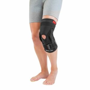

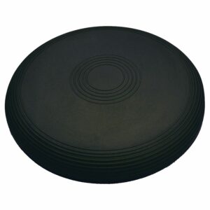
To purchase physiotherapy products to assist with rehabilitation click on one of the above links or visit the PhysioAdvisor Shop.

Link to this Page
If you would like to link to this article on your website, simply copy the code below and add it to your page:
<a href="https://physioadvisor.com.au/injuries/knee”>Diagnostic Guide – Knee Pain – PhysioAdvisor.com</a><br/>PhysioAdvisor provides a diagnostic guide for patients suffering from knee pain and knee injuries created by experienced physiotherapists.
Return to the top of Diagnostic Guide – Knee Pain.