Carpal Bone Anatomy: Bony Landmarks, Muscular Attachments, and Common Injuries
Updated:
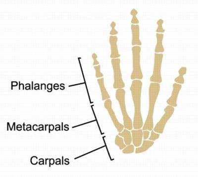
Learning carpal bone anatomy is a crucial aspect of understanding the structure and function of the human wrist and hand. The carpal bones are small, irregularly shaped bones located in the wrist joint, which connect the forearm to the hand. These bones work together with the radius and ulna bones of the forearm to provide flexibility and stability to the hand and wrist.
In this article, we will discuss the bony landmarks, muscular attachments, and common injuries associated with carpal bone anatomy.
Carpal Bone Anatomy – Bony Landmarks
The eight carpal bones can be divided into two rows: the proximal row and the distal row. The proximal row is located closer to the forearm, while the distal row is closer to the hand.
The proximal row consists of the scaphoid, lunate, triquetral, and pisiform bones. The distal row consists of the trapezium, trapezoid, capitate, and hamate bones.
Each bone has specific bony landmarks that are important for identifying and diagnosing injuries. For example, the scaphoid bone has a tubercle on the lateral side, which is a common site of injury. The capitate bone has a prominent head that is palpable on the back of the hand, while the hamate bone has a hook-like process on its palmar surface.
Carpal Bone Anatomy – Muscular Attachments
The carpal bones provide attachment sites for several muscles that move the hand and wrist. The flexor carpi radialis muscle attaches to the base of the second metacarpal bone and the anterior surface of the radial styloid process, while the flexor carpi ulnaris muscle attaches to the pisiform bone and the hook of the hamate bone.
The extensor carpi radialis muscle attaches to the base of the second and third metacarpal bones and the posterior surface of the lateral epicondyle of the humerus, while the extensor carpi ulnaris muscle attaches to the base of the fifth metacarpal bone and the posterior surface of the ulna.
Common Injuries of Carpal Bones
Injuries to the carpal bones can be caused by a variety of factors, including trauma, overuse, and degenerative conditions. Some of the most common injuries associated with carpal bones include:
- Scaphoid fractures: Fractures of the scaphoid bone are common and can be caused by falls onto an outstretched hand. These fractures can be difficult to diagnose and may require imaging studies such as X-rays or MRI.
- Triquetral fractures: Fractures of the triquetral bone are less common than scaphoid fractures but can also be caused by falls onto an outstretched hand. These fractures are usually diagnosed with X-rays.
- Hamate fractures: Fractures of the hook of the hamate bone are often associated with sports that involve swinging a racket or club. These fractures can be diagnosed with X-rays or MRI.
- Kienbock’s disease: This is a degenerative condition that affects the lunate bone and is caused by a disruption of its blood supply. Symptoms may include pain, stiffness, and decreased grip strength.
- Other injuries affecting the tissue surrounding carpal bones are also common such as wrist sprains, wrist tendinopathy, carpal tunnel syndrome.
Conclusion
Understanding carpal bone anatomy is essential for diagnosing and treating injuries and conditions that affect the hand and wrist. By knowing the bony landmarks and muscular attachments of the carpal bones, healthcare professionals can identify and treat injuries effectively. With this knowledge, patients can receive optimal care and achieve the best possible outcomes.
References
- Bones of the Hand: Carpal Bone Anatomy by Teach Me Anatomy: https://teachmeanatomy.info/upper-limb/bones/bones-of-the-hand-carpals-metacarpals-and-phalanges/
- Moore, K. L., Dalley, A. F., & Agur, A. M. (2013). Clinically oriented anatomy. Lippincott Williams & Wilkins.
- Netter, F. H. (2014). Atlas of human anatomy. Elsevier Health Sciences.
- Standring, S. (Ed.). (2016). Gray’s anatomy: the anatomical basis of clinical practice. Elsevier Health Sciences.
- Placzek, J. D., Boyer, M. I., & Calfee, R. P. (2010). Acute carpal injuries: diagnosis and management. Journal of the American Academy of Orthopaedic Surgeons, 18(6), 328-336.
- Trumble, T. E., & Gilbert, M. (2017). Carpal injuries: diagnosis and management. Springer.

Link to this Page
If you would like to link to this article on your website, simply copy the code below and add it to your page:
<a href="https://physioadvisor.com.au/health/anatomy/bones/carpal-bone-anatomy”>Carpal Bone Anatomy: Bony Landmarks, Muscular Attachments, and Common Injuries – PhysioAdvisor.com</a><br/>Learn about carpal bone anatomy including bony landmarks, muscular attachements and common injuries on PhysioAdvisor.
Return to the top of Carpal Bone Anatomy: Bony Landmarks, Muscular Attachments, and Common Injuries.