Cervical Vertebrae
Updated:
Relevant Bony Anatomy
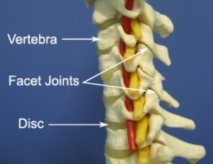
The cervical vertebrae are the smallest of the moving vertebrae of the spine. There are seven in total (C1 to C7) which form the neck and are numbered from the top of the neck (C1) to the bottom (C7). Collectively they act to support the skull, protect the spinal cord, allow movement of the neck and provide attachment points for the muscles and ligaments of the neck. Each cervical vertebrae joins with adjacent vertebrae primarily at the facet joints (located on each side of the spine) and the discs of the neck (located centrally at the front of the spine). Movements between each adjacent vertebrae are relatively small, but when summated over the entire vertebral column allow considerable mobility.
Apart from C1 and C2 which are atypical cervical vertebrae due to their structure and unique function (i.e. C1 attaches to the skull), the remaining cervical vertebrae primarily comprise of a body (at the front of the bone) and a vertebral arch (situated directly behind the body) which forms a relatively large hole known as the vertebral foramen. Since each vertebrae is situated directly above or below each other, their collective vertebral foramen line up forming the vertebral canal which houses and protects the spinal cord. Each vertebrae also has various bony prominences, such as the spinous process (located at the back of the bone) and transverse processes (located at each side of the vertebrae). These bony prominences provide attachment points to the ligaments and muscles of the neck.

Figure 1 – Anatomy of the Cervical Vertebrae & Neck
Location
The seven cervical vertebrae are located directly beneath the skull and are numbered from the top to bottom – C1 to C7.
Forms Joints With
- The Skull – C1 (also known as the Atlas) forms a joint with the skull above and C2 below.
- Each of the remaining vertebrae of the neck (C2 – C7) connect with the vertebrae above and below via bony processes on each side of the spine which form the facet joints (figure 1) and via the discs located centrally between each spinal segment (figure 1).
- T1 (first thoracic vertebre) – C7 also forms joints with the first thoracic vertebrae (T1) via the facet joints and discs.
Major Muscles of the Neck
- Cervical Erector Spinae – Muscle group at the back of the neck responsible for keeping the spine erect, bending the neck backwards and assisting side bending and rotation movements located on each side of the body.
- Splenius Capitis – Muscle located at the back of the neck on each side of the body acting to side bend, rotate and arch the neck backwards.
- Deep Cervical Flexors – Muscle group located deep within the front of the neck responsible for helping to bend the neck forwards and stabilising the joints of the neck.
- Levator Scapulae – Muscle attaching the top of the shoulder blade to the side of the neck, responsible for elevating the shoulder blade (scapula) and side bending the neck.
- Scalenes – Muscle group located on each side of the neck, responsible for bending the neck to the side and elevating the first two ribs during respiration.
- Sternocleidomastoid – Muscle at the front of the neck located on each side of the midline attaching the collar bone and breast bone to the mastoid process of the skull just behind the lower aspect of the ear.
- Platysma – Superficial muscle located at the front of the neck on each side of the midline of the body.
- Hyoid Muscles – A group of muscles located at the front of the neck primarily responsible for moving the hyoid bone and larynx which aids in tongue movements, speech and swallowing.
Other Attachments
- With the exception of C1, each cervical vertebrae connects with the vertebrae above and below via an intervertebral disc.
- Numerous strong ligaments reinforce connections between adjacent vertebrae and discs, providing stability.
Related injuries
- Cervical Disc Bulge
- Facet Joint Sprain
- Cervicogenic Headache
- Neck Arthritis
- Whiplash
- Postural Syndrome
- Wry Neck (Discogenic)
- Wry Neck (Facet)
- Fractures
Relevant Physiotherapy Exercises
Recommended Reading
- View our Head and Neck Diagnosis Guide.
- View detailed information on improving your Posture.
- View detailed information on Postural Taping.
- View detailed information on Ergonomic Computer Setup.
- View detailed information on Mobile Phone Ergonomics.
- View detailed information on Choosing a School Bag.
- View detailed information on Safe Lifting.
Find a Physio
Find a physiotherapist in your local area who can diagnose and treat sports and spinal injuries and provide education on the anatomy of the neck and cervical vertebrae.

Link to this Page
If you would like to link to this article on your website, simply copy the code below and add it to your page:
<a href="https://physioadvisor.com.au/health/anatomy/bones/cervical-vertebrae”>Cervical Vertebrae – PhysioAdvisor.com</a><br/>PhysioAdvisor provides detailed physiotherapy information on the human anatomy of the neck and cervical vertebrae. Including location, joints, muscular attachments, relevant injuries and more...
Return to the top of Cervical Vertebrae.


