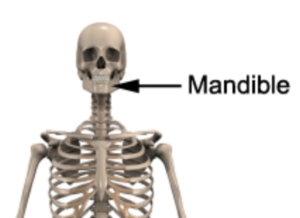
Mandible
Updated:
Relevant Bony Anatomy
The mandible (jawbone) is the bony structure of the lower jaw and lowest aspect of the face. It is the largest and strongest of the facial bones and provides sockets (known as alveolar processes) to hold the lower teeth in place. The mandible primarily acts to support the lower aspect of the face, provide a fixation point for the lower teeth, allow jaw movement during speech and mastication (chewing) and is essential for other movements of the mouth (such as yawning).
The mandible forms the lower aspect of the skull and consists of two parts: a horizontal part called the body, and two vertical parts called rami which ascend almost vertically from the back of the mandibular body. The top of each ramus contains two bony prominences known as the condylar and coronoid processes.

Figure 1 – Relevant Anatomy of the Mandible
Location
The mandible is located at the bottom aspect of the skull and forms the lower jaw bone (figure 1).
Forms Joints With
The mandible forms joints with the temporal bone of the cranium at the Temporo-Mandibular Joint (TMJ)
Major Muscular Attachments
- Muscles of facial expression
- Chewing Muscles:
- Masseter – this quadrangular chewing muscle attaches from the side of the mandible’s ramus on each side of the jaw to the skull above (i.e. the zygomatic arch). The masseter elevates and protrudes the mandible, thereby closing the jaw. Deeper fibres of the muscle retrude the jaw.
- Temporalis – This extensive and powerful fan shaped chewing muscle originates from the temporal bone of the skull and attaches to the ramus of the mandible and its coronoid process. The temporalis muscle elevates the mandible, closing the jaws. It’s posterior muscle fibres retrude the mandible after protrusion.
- Lateral Pterygoid – this muscle has a conical appearance originating from the lower aspect of the temporal bone and sphenoid bones of the skull and attaching to the neck of the mandible as well as the capsule and articular disc of the temporo-mandibular joint. Acting alone, the lateral pterygoid muscles produce side to side movements of the mandible. Acting together they protrude the mandible and depress the chin.
- Medial Pterygoid – This quadrilateral muscle originates from the skull (pterygoid plate, palatine bone and tuberosity of the maxilla) and inserts into the inner surface of the ramus of the mandible. The medial pterygoid muscles help to elevate the mandible, closing the jaws. Acting together, they help to protrude the mandible. Acting alone, it protrudes the side of the jaw and can produce a grinding motion when acting alternately.
Related injuries
- Cervicogenic Headache
- TMJ Dysfunction
- Fractures
Relevant Physiotherapy Exercises
Recommended Reading
- View our Head and Neck Diagnosis Guide.
- View detailed information on improving your Posture.
- View detailed information on Postural Taping.

Find a Physio
Find a physiotherapist in your local area who can diagnose and treat sports and spinal injuries and provide education on the anatomy of the mandible and skull.

Link to this Page
If you would like to link to this article on your website, simply copy the code below and add it to your page:
<a href="https://physioadvisor.com.au/health/anatomy/bones/mandible”>Mandible – PhysioAdvisor.com</a><br/>PhysioAdvisor provides detailed physiotherapy information on the human anatomy of the mandible (lower jaw). Including location, joints, muscular attachments, relevant injuries and more...
Return to the top of Mandible.


