Skull
Updated:
Relevant Bony Anatomy
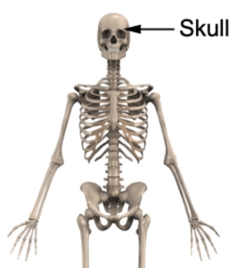
The skull is the bony structure of the head that primarily acts to protect the brain, sense organs and top of the spinal cord whilst providing support for the structures of the face. It is one of the most complex bony structures of the human skeleton as it encloses the brain, contains the organs of senses for hearing, seeing, tasting and smelling and provides an opening for the digestive and respiratory tracts.
The skull is made up of 22 separate bones that are closely fitted together and mostly joined by immovable fibrous joints known as sutures. In babies, there are relatively large gaps between the individual bones, giving the head malleability and allowing it to squeeze through the birth canal. These gaps present in babies are called fontanelles and are covered by fibrous membranes. The skull as a whole tends to be of greater clinical importance than its individual, constituent bones and can be divided into two main sections – the cranium (comprising mostly of strong immovable fibrous joints between adjacent bones) and the mandible (lower jaw).

Figure 1 – Relevant Bony Anatomy of the Skull
Location
The skull is located directly above the neck and is situated at the top of the axial skeleton during standing (figure 1).
Forms Joints With
The skull has numerous, strong, fibrous joints between many of its 22 constituent bones. As a whole, the cranium also forms joints with the following bones:
- C1 (Atlas) – First cervical vertebra (neck)
- The mandible (jaw) – i.e. the mandible forms a joint with the temporal bone of the cranium at the Temporo-Mandibular Joint (TMJ)
Major Muscular Attachments
- Muscles of facial expression
- Chewing Muscles – Masseter, Temporalis and Pterygoids.
- Erector Spinae – Muscle group at the back of the neck responsible for keeping the spine erect, located on each side of the body and attaching to the back of the cranium.
- Trapezius – Muscle located between the shoulder blade and neck, attaching the top of the shoulder blade (scapula) to the occipital bone at the base of the back of the cranium on each side of the body.
- Platysma – Superficial muscle located at the front of the neck on each side of the midline of the body.
- Sternocleidomastoid – Muscle at the front of the neck located on each side of the midline attaching the collar bone and breast bone to the mastoid process of the cranium just behind the lower aspect of the ear.
Related injuries
- Cervicogenic Headache
- Whiplash
- Postural Syndrome
- Neck Arthritis
- TMJ Dysfunction
- Fractures
- Concussion
- Traumatic Brain Injury
Relevant Physiotherapy Exercises
More Information
- View our Head and Neck Diagnosis Guide.
- View detailed information on improving your Posture.
Find a Physio
Find a physiotherapist in your local area who can diagnose and treat sports and spinal injuries and provide education on the anatomy of the skull and cranium.

Link to this Page
If you would like to link to this article on your website, simply copy the code below and add it to your page:
<a href="https://physioadvisor.com.au/health/anatomy/bones/skull”>Skull – PhysioAdvisor.com</a><br/>PhysioAdvisor provides detailed physiotherapy information on the human anatomy of the skull and cranium. Including location, joints, muscular attachments, relevant injuries and more...
Return to the top of Skull.


