Metacarpal Anatomy: Understanding the Bones of Your Hand
Updated:
If you have ever wondered about the intricate workings of your hand, you are not alone. The hand is a marvel of biomechanical engineering, and it is essential for everything from daily tasks to athletic performance. At the heart of the hand are the five metacarpal bones. In this article, we will explore the anatomy of these bones, their muscular attachments, common injuries, and bony landmarks.
Bony Landmarks
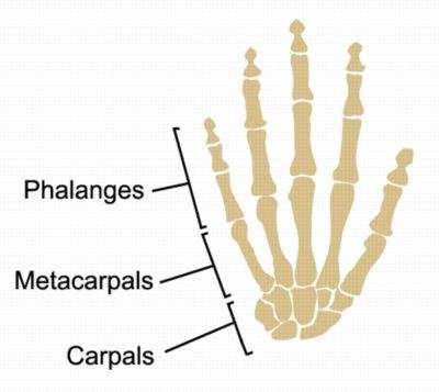
The metacarpals are long bones that connect the wrist to the fingers. Each metacarpal has a base, shaft, and head. The base of the metacarpal bone connects to the bones of the wrist (carpals), while the head connects to the bones of the fingers (Phalanges – Figure 1). The shaft is the middle portion of the bone.
The metacarpals are numbered one through five, starting with the thumb side. The first metacarpal is the shortest and thickest, while the fifth metacarpal is the thinnest. The metacarpals also have various bony landmarks that serve as attachment points for muscles and ligaments.
Metacarpal Anatomy: Muscular Attachments
There are many muscles that attach to the metacarpals. The muscles that control movement of the fingers and thumb originate from the forearm and attach to the fingers via tendons that run over the wrist and across the palm.
The muscles and their respective tendons that attach to the metacarpals (and those that travel adjacent to them) are responsible for movement of the fingers and thumb. For example, the interossei muscles are intrinsic muscles that lie between the metacarpal bones and help to control sideways movement of the fingers (abduction – moving the fingers apart and adduction bringing them together). The flexor digitorum profundus muscle originates from the forearm and attaches to the distal phalanges of the fingers. This muscle is responsible for flexing the fingers. Other muscles, such as the extensor digitorum, are responsible for extending the fingers.
Metacarpal Anatomy: Common Injuries
Metacarpal fractures are common injuries that occur when there is a direct blow to the hand, such as during a fall or when hitting a hard object. The most common type of metacarpal fracture is known as a “boxer’s fracture,” which occurs when the fifth metacarpal is fractured due to a punch or other direct impact.
Other injuries involving damage to soft tissue connecting to the metacarpals include dislocations, sprains, and ligament tears. These injuries can occur due to repetitive motion or trauma, such as during sports or other physical activities.
Conclusion
In conclusion, understanding the anatomy of the metacarpal bones is important for anyone interested in the function and health of their hands. By knowing the bony landmarks, muscular attachments, and common injuries, you can better care for your hands and help to identify potential injuries. If you have any concerns about your hand health, be sure to consult with a qualified Physiotherapist.
References
- Moore KL, Dalley AF. Clinically Oriented Anatomy. 7th ed. Philadelphia, PA: Lippincott Williams & Wilkins; 2014.
- Drake RL, Vogl AW, Mitchell AW. Gray’s Anatomy for Students. 2nd ed. Philadelphia, PA: Churchill Livingstone/Elsevier; 2010.
- Rettig AC. Athletic Training and Sports Medicine: An Integrated Approach. 5th ed. Burlington, MA: Jones & Bartlett Learning; 2017.
- Metacarpal Bones by Wikipedia: Metacarpal bones – Wikipedia

Link to this Page
If you would like to link to this article on your website, simply copy the code below and add it to your page:
<a href="https://physioadvisor.com.au/metacarpal-anatomy”>Metacarpal Anatomy: Understanding the Bones of Your Hand – PhysioAdvisor.com</a><br/>Learn about Metacarpal Anatomy including bony landmarks, muscular attachments and common injuries on PhysioAdvisor.
Return to the top of Metacarpal Anatomy: Understanding the Bones of Your Hand.