Metatarsal Anatomy: Understanding the Long Bones of the Foot
Updated:
The foot is a complex structure made up of numerous bones, muscles, and ligaments that work together to support our body weight and allow us to move around. In this article, we will explore metatarsal anatomy, which are the bones located in the middle of the foot, including their bony landmarks, muscular attachments, common injuries, and treatments.
Metatarsal Anatomy – Bony Landmarks
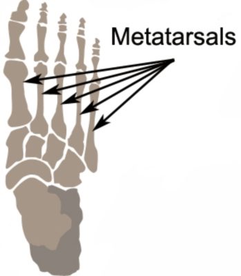
The metatarsals are five long bones that extend from the tarsal bones in the ankle to the phalanges in the toes. They are numbered from one to five, starting with the first metatarsal on the inside of the foot and ending with the fifth metatarsal on the outside. Each metatarsal has a base, a shaft, and a head, and is connected to the adjacent metatarsals by small joints called intermetatarsal joints.
Metatarsal Anatomy – Muscular Attachments
Several muscles attach to the metatarsals, including the muscles of the foot and ankle, such as the flexor hallucis brevis, flexor digitorum brevis, and abductor hallucis muscles. These muscles work together to control the movement of the foot and toes, and to provide support to the arch of the foot.
Common Injuries affecting the Metatarsals
The metatarsals are susceptible to a variety of injuries, including metatarsal stress fractures, which occur due to repetitive overuse or sudden impact. These types of injuries are common among athletes who engage in activities that require a lot of running or jumping, such as basketball, soccer, or track and field.
Another common injury is a metatarsal fracture, which can occur due to a sudden impact, such as a fall or a collision. These types of injuries are also common among athletes, but can occur in anyone who experiences trauma to the foot.
Treatment for metatarsal injuries may include rest, ice, compression, and elevation (RICE), physical therapy, and in some cases, surgery. Physical therapy can be particularly helpful for restoring foot strength and mobility after an injury or surgery.
References – Metatarsal Anatomy
- Bones of the Foot – Tarsals, Metatarsals and Phalanges by Teach me Anatomy – Bones of the Foot – Tarsals – Metatarsals – Phalanges – TeachMeAnatomy
- Moore KL, Dalley AF. Clinically Oriented Anatomy. 7th ed. Philadelphia, PA: Lippincott Williams & Wilkins; 2014.
- Drake RL, Vogl AW, Mitchell AW. Gray’s Anatomy for Students. 2nd ed. Philadelphia, PA: Churchill Livingstone/Elsevier; 2010.
- Magee DJ. Orthopedic Physical Assessment. 6th ed. St. Louis, MO: Elsevier Saunders; 2014.
In conclusion, the metatarsals are an essential part of the foot’s structure and function. By understanding the bony landmarks, muscular attachments, and common injuries associated with the metatarsals, patients can better care for their feet and prevent injuries. If you have any concerns about your foot health, be sure to consult with a qualified healthcare provider.

Link to this Page
If you would like to link to this article on your website, simply copy the code below and add it to your page:
<a href="https://physioadvisor.com.au/metatarsal-anatomy”>Metatarsal Anatomy: Understanding the Long Bones of the Foot – PhysioAdvisor.com</a><br/>Learn about metatarsal anatomy on PhysioAdvisor including bony landmarks, muscular attachments, common injuries and more...
Return to the top of Metatarsal Anatomy: Understanding the Long Bones of the Foot.