Patella Anatomy: Understanding the Kneecap
Updated:
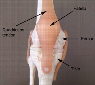
The patella, or kneecap, is a small bone located at the front of the knee joint in the patellofemoral groove of the femur (figure 1). It is a key component of the knee joint and plays an important role in knee movement and stability. In this article, we will explore the anatomy of the patella, including its bony landmarks, muscular attachments, common injuries, and treatments.
Patella Anatomy: Bony Landmarks
The patella is a flat, triangular bone that sits in the tendon of the quadriceps muscle (figure 1). It has a base, an apex, and two articular surfaces that connect with the femur bone at the patello-femoral joint. The base of the patella is the widest part of the bone, and the apex is the thinnest part.
The patella also has several bony landmarks that serve as attachment points for muscles and ligaments. The patellar ligament attaches to the inferior pole (bottom section) of the patella, while the quadriceps tendon attaches to the superior pole (top section). The medial and lateral patellar retinacula (fibrous connective tissue located at the inner and outer parts of the patella) are also attachment points for ligaments and muscles.
Patella Anatomy: Muscular Attachments
Several muscles attach to the patella, including the quadriceps muscle, which is responsible for extending the knee joint. The quadriceps muscle is the largest muscle in the body and has four separate muscle bellies that all merge into one common tendon, which surrounds the patella and attaches to the tibia bone in the lower leg.
The four separate muscle bellies of the quadriceps that attach to the patella include the vastus medialis, vastus lateralis, rectus femoris and vastus intermedius muscle. The articularis genu muscle also attaches to the patella. These muscles play important roles in knee movement and stability.
Common Injuries affecting the Patella
The patella is susceptible to a variety of injuries, including patella fractures, patella dislocations, patellofemoral pain syndrome and patellar tendonitis. Patellar tendonitis, also known as jumper’s knee, is a common injury that occurs when the patellar tendon becomes inflamed due to repetitive jumping or other activities that place stress on the knee.
Patellar dislocations occur when the patella is forced out of its normal position, often due to a sudden twisting motion or trauma. Fractures affecting the patella can also occur due to a direct blow to the knee or as a result of a fall.
Treatment for patella injuries may include rest, ice, compression, and elevation (RICE), physiotherapy, and in some cases, surgery. Physiotherapy is essential to ensure an optimal outcome in all conditions and can be particularly helpful for restoring knee strength and mobility after an injury or surgery.
Conclusion
In conclusion, the patella is an important bone in the knee joint that plays a crucial role in knee movement and stability. By understanding the bony landmarks, muscular attachments, and common injuries associated with the patella, patients can better care for their knees and address any injuries that may arise. If you have any concerns about your knee health, be sure to consult with a qualified Physiotherapist.
Patella Anatomy – References
- Patella Anatomy by Teach Me Anatomy – The Patella – Surface Anatomy – Functions – Dislocation – TeachMeAnatomy
- Moore KL, Dalley AF. Clinically Oriented Anatomy. 7th ed. Philadelphia, PA: Lippincott Williams & Wilkins; 2014.
- Drake RL, Vogl AW, Mitchell AW. Gray’s Anatomy for Students. 2nd ed. Philadelphia, PA: Churchill Livingstone/Elsevier; 2010.
- Magee DJ. Orthopedic Physical Assessment. 6th ed. St. Louis, MO: Elsevier Saunders; 2014.

Link to this Page
If you would like to link to this article on your website, simply copy the code below and add it to your page:
<a href="https://physioadvisor.com.au/patella-anatomy”>Patella Anatomy: Understanding the Kneecap – PhysioAdvisor.com</a><br/>Learn about patella anatomy on PhysioAdvisor including bony landmarks, muscular attachments and common injuries.
Return to the top of Patella Anatomy: Understanding the Kneecap.