Pectoralis Minor Anatomy
Updated:
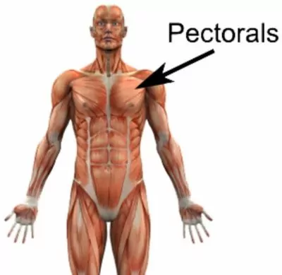
The pectoralis minor muscle is a crucial component of the anatomy of the chest, playing a significant role in shoulder stability and movement. In this article, we will delve into the intricacies of pectoralis minor anatomy, exploring its origin, insertion, action, nerve supply, blood supply, common injuries, and more. By understanding the intricacies of this muscle, we can gain insights into its functions and potential issues. Let’s dive in!
Origin:
The pectoralis minor originates from the anterior surfaces of the third, fourth, and fifth ribs near their costal cartilages. It also has attachments to the aponeurosis of the external oblique muscle, which further contributes to its origin.
Insertion:
The pectoralis minor inserts onto the coracoid process of the scapula, a bony prominence located on the anterior surface of the shoulder blade. This insertion point allows the muscle to exert control and influence over the position and movement of the scapula.
Action:
The primary action of the pectoralis minor muscle is to depress the scapula. This downward movement of the shoulder blade assists in stabilizing the shoulder joint, especially during activities that involve reaching overhead or pushing movements. Additionally, the pectoralis minor muscle contributes to protraction (forward movement) and downward rotation of the scapula.
Nerve Supply:
The pectoralis minor muscle is innervated by the medial pectoral nerve, which arises from the brachial plexus. This nerve originates from the ventral rami of the spinal nerves C8 and T1.
Blood Supply:
The blood supply to the pectoralis minor muscle is primarily derived from the pectoral branch of the thoracoacromial artery, a branch of the axillary artery. This arterial supply ensures the delivery of oxygen and nutrients necessary for the muscle’s proper functioning.
Common Injuries:
- Pectoralis Minor Strain: Overuse or sudden trauma can result in a strain or tear of the pectoralis minor muscle. Symptoms may include pain, weakness, and restricted shoulder movement. Rest, ice, compression, and physical therapy are commonly employed for rehabilitation.
- Thoracic Outlet Syndrome: In some cases, compression of the neurovascular bundle passing beneath the pectoralis minor muscle can occur, leading to thoracic outlet syndrome. This condition can cause pain, numbness, and tingling sensations in the arm and hand. Treatment options include physical therapy, medications, and, in severe cases, surgery.
Pectoralis Minor Anatomy – Conclusion
The pectoralis minor muscle is an important component of the chest anatomy, contributing to shoulder stability and movement. Understanding its origin, insertion, action, nerve supply, and blood supply provides insights into its role in maintaining optimal shoulder function. Awareness of common injuries associated with this muscle can help individuals seek timely treatment and rehabilitation when needed. Stay proactive in maintaining shoulder health by incorporating targeted exercises and seeking professional advice if you experience any discomfort or pain.
Pectoralis Minor Anatomy – References:
- Netter, F. H. (2014). Atlas of human anatomy. Elsevier Health Sciences.
- Drake, R. L., Vogl, W., Mitchell, A. W. M., & Gray, H. (2010). Gray’s anatomy for students. Elsevier Health Sciences.
- Physiopedia: Pectoralis Minor – Physiopedia (physio-pedia.com)

Link to this Page
If you would like to link to this article on your website, simply copy the code below and add it to your page:
<a href="https://physioadvisor.com.au/pectoralis-minor-anatomy”>Pectoralis Minor Anatomy – PhysioAdvisor.com</a><br/>Learn about pectoralis minor anatomy on PhysioAdvisor including origin, insertion, blood and nerve supply, actions, common injuries and more.
Return to the top of Pectoralis Minor Anatomy.