Pelvis Anatomy
Updated:
The pelvis is a complex structure that plays a crucial role in the foundation of the human skeleton. It provides support for the spine, lower extremities, and internal organs. Understanding the anatomy of the pelvis is essential for diagnosing and treating injuries and conditions related to this area. In this article, we will discuss the bony landmarks, muscular attachments, and common injuries associated with the pelvis.
Bony Landmarks
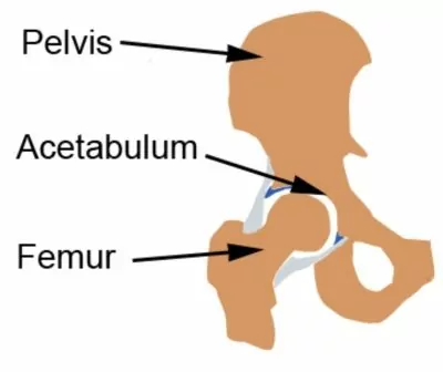
The pelvis is composed of three bones: the ilium, ischium, and pubis. These bones come together to form the acetabulum (figure 1), which is the socket of the hip joint. The ilium is the largest of the three bones and forms the upper portion of the pelvis. It has several important landmarks, including the iliac crest, which is the ridge at the top of the hip bone, and the greater sciatic notch, which is a large notch on the back of the ilium. The ischium is the bone that supports the weight of the body when sitting and has a bony prominence called the ischial tuberosity (i.e.sitting bone). The pubis is the bone that forms the front of the pelvis and has a pubic symphysis, which is the joint where the two pubic bones meet.

Muscular Attachments
The pelvis has numerous muscular attachments that allow for movement and stability. The gluteus maximus muscle attaches to the ilium and ischium and allows for extension (i.e. backward movement) of the hip joint. The adductor muscles attach to the pubis and ischium and allow for adduction (movement towards the midline of the body) of the hip joint. The hip flexor muscles, including the iliopsoas and rectus femoris, attach to the ilium and allow for flexion of the hip joint (i.e. knee toward chest movement).
Pelvis Anatomy – Common Injuries
Injuries to the pelvis at times can be severe and potentially life-threatening. Fractures, dislocations, and sprains are some injuries that may occur, associated with the pelvis. Pelvic fractures can occur from high-impact trauma, such as a car accident or fall from a height, and can result in significant pain, swelling, and immobility. Pelvic Stress Fractures are typically associated with overuse. Dislocations of the hip joint can occur from a traumatic event (e.g. motor vehicle accident) and can cause severe pain and difficulty with movement. Sprains, which involve the tearing or stretching of ligaments, can occur from overuse or trauma and can result in pain and instability. Overuse injuries to tendons attaching to the pelvis can also occur such as adductor tendinopathy or hamstring tendinopathy. Osteitis Pubis and trochanteric bursitis are other common injuries associated with the pelvis.
Understanding the anatomy of the pelvis is crucial for identifying and treating injuries and conditions related to this area. The bony landmarks, muscular attachments, and common injuries associated with the pelvis provide a framework for healthcare professionals to diagnose and treat these injuries. If you experience pain or discomfort in your pelvic region, it is important to consult with a healthcare professional to determine the cause and best course of treatment.
Pelvis Anatomy – References:
- Moore, K. L., Dalley, A. F., & Agur, A. M. (2013). Clinically oriented anatomy. Lippincott Williams & Wilkins.
- Netter, F. H. (2014). Atlas of human anatomy. Elsevier Health Sciences.
- Standring, S. (Ed.). (2016). Gray’s anatomy: the anatomical basis of clinical practice. Elsevier Health Sciences.
- Ponsford, F. M., & Hill, A. M. (2016). Pelvic fractures in the elderly: a review. EFORT Open Reviews, 1(7), 275-281. doi: 10.1302/2058-5241.1.000053
- Kohn, M. A., & Carpenter, C. R. (2012). Evaluation of acute hip pain in adults. American Family Physician, 86(4), 350-356.
- The Pelvis by teach me anatomy : The Pelvis – TeachMeAnatomy

Link to this Page
If you would like to link to this article on your website, simply copy the code below and add it to your page:
<a href="https://physioadvisor.com.au/pelvis-anatomy”>Pelvis Anatomy – PhysioAdvisor.com</a><br/>Learn about pelvis anatomy on PhysioAdvisor including bony landmarks, muscular attachments and common injuries.
Return to the top of Pelvis Anatomy.
