Radius Anatomy: Structure and Function
Updated:
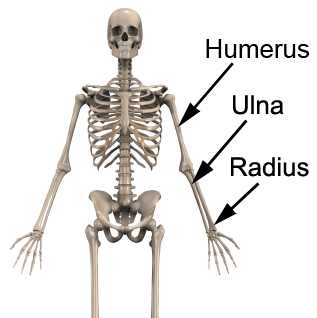
The radius is a long bone in the forearm that extends from the elbow to the wrist, forming the lateral (outer) border of the forearm. It is one of the two bones in the forearm, the other being the ulna, and plays a critical role in the function and mobility of the wrist and hand. In this article, we will explore the anatomy of the radius, including its bony landmarks, muscular attachments, and related injuries.
Radius Anatomy
Bony Landmarks of the Radius
The radius has two ends and a shaft. The proximal end of the radius is the upper end that articulates with the ulna and humerus at the elbow joint. The distal end of the radius is the lower end that articulates with the bones of the wrist, including the scaphoid, lunate, and triquetrum. The shaft of the radius is curved and has a smooth surface, making it one of the strongest and most resilient bones in the body.
The radius also has several bony landmarks that provide attachment sites for ligaments and muscles. The radial tuberosity is a small, anterior prominence on the proximal end of the radius that serves as the attachment site for the biceps brachii muscle. The styloid process is a small, pointed projection on the distal end of the radius that serves as the attachment site for the wrist ligaments.
Radius Anatomy: Muscular Attachments
The radius has several muscular attachments that are important for the movement and stability of the wrist and hand. The biceps brachii muscle attaches to the radial tuberosity of the radius and helps to flex the elbow joint. The pronator teres muscle attaches to the proximal end of the radius and helps to rotate the forearm inward (pronation). The supinator muscle attaches to the proximal end of the radius and helps to rotate the forearm outwards (supination).
Injuries of the Radius
The radius is susceptible to various types of injuries due to its function and location. Fractures of the radius can occur due to falls, direct trauma, or overuse injuries. These fractures can cause pain, swelling, and deformity of the wrist, requiring medical attention. Dislocations of the wrist joint can also cause damage to the radius, causing pain and limited range of motion. Arthritis and tendonitis (of the relevant joints / muscles respectively) can also affect the radius, causing pain and inflammation. Occasionally, damage to the radioulnar joint can also occur such as sprains or dislocations.
Conclusion
The radius is a crucial bone that plays a pivotal role in the stability and mobility of the wrist and hand. Its bony landmarks and muscular attachments allow for a wide range of movement, while its resilience and strength protect it from injuries. Understanding the anatomy of the radius can help to prevent and manage injuries, allowing for optimal function and performance of the wrist and hand joints.
Radius Anatomy References:
“Radius Anatomy” by Healthline (https://www.healthline.com/human-body-maps/radius-bone#1)
“Anatomy of the Radius” by Teach Me Anatomy (https://teachmeanatomy.info/upper-limb/bones/radius/)

Link to this Page
If you would like to link to this article on your website, simply copy the code below and add it to your page:
<a href="https://physioadvisor.com.au/radius-anatomy”>Radius Anatomy: Structure and Function – PhysioAdvisor.com</a><br/>Learn about radius anatomy including bony landmarks, muscular attachments and common injuries on PhysioAdvisor
Return to the top of Radius Anatomy: Structure and Function.