Exploring Subscapularis Anatomy: Function, Injuries, and Clinical Relevance
Updated:
The subscapularis muscle is a vital component of the shoulder complex, playing a crucial role in stabilizing and mobilizing the shoulder joint. In this article, we will delve into the intricate details of subscapularis anatomy, its origin, insertion, action, nerve supply, blood supply, palpation techniques, clinical relevance, and common injuries.
Subscapularis Anatomy:
Origin and Insertion:
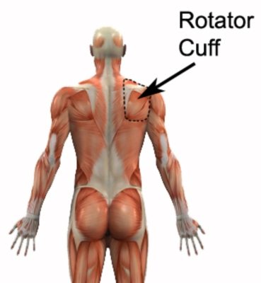
The subscapularis muscle is one of the four muscles that make up the rotator cuff in the shoulder. It originates from the subscapular fossa, a concave depression on the anterior (front) surface of the scapula (shoulder blade). From this point of origin, the muscle fibers converge to form a tendon, which inserts onto the lesser tubercle of the humerus, located on the front of the upper arm bone.
Action and Function:
The primary action of the subscapularis muscle is internal rotation of the humerus. This means that it helps to turn the upper arm bone inward toward the body’s midline. Additionally, the subscapularis assists in the stabilization of the shoulder joint, preventing dislocation during arm movements. It works in harmony with the other rotator cuff muscles to ensure the smooth functioning of the shoulder.
Nerve Supply:
The subscapularis muscle receives its nerve supply from the upper and lower subscapular nerves, which are branches of the brachial plexus. These nerves play a pivotal role in transmitting signals from the brain to the muscle, enabling it to contract and perform its functions effectively.
Blood Supply:
The subscapularis muscle is predominantly supplied by the subscapular artery, a branch of the axillary artery. A robust blood supply is essential for providing oxygen and nutrients to the muscle, aiding in its function and recovery.
Palpation of Subscapularis Anatomy:
Palpating the subscapularis muscle can be challenging due to its location deep within the shoulder. To palpate it, you can place your hand on the anterior aspect of the shoulder and gently press inward while asking the patient to resist your pressure as they perform internal rotation. This allows you to feel the muscle contracting beneath your fingers.
Subscapularis Anatomy – Clinical Relevance:
The subscapularis muscle is of significant clinical importance, particularly in the fields of orthopedics and Physiotherapy. Understanding its anatomy and function is crucial for diagnosing and treating various shoulder conditions and injuries.
Common Injuries related to Subscapularis:
- Subscapularis Tears: These tears can occur due to trauma or overuse, leading to pain, weakness, and limited internal rotation of the shoulder.
- Rotator Cuff Impingement: Subscapularis involvement in rotator cuff impingement can cause pain and discomfort when raising the arm, particularly during overhead activities.
- Shoulder Instability: A weakened subscapularis can contribute to shoulder instability, increasing the risk of dislocation.
- Frozen Shoulder (Adhesive Capsulitis): This condition can lead to stiffness and limited mobility in the shoulder, with the subscapularis often affected.
- Rehabilitation: Proper rehabilitation of subscapularis injuries is essential to restore function and prevent long-term issues. Physiotherapists often employ exercises targeting this muscle to aid recovery.
Subscapularis Anatomy – Useful Links:
For more information on subscapularis anatomy and related topics, you can explore the following articles on PhysioAdvisor:
- Rotator Cuff Tears – PhysioAdvisor
- Shoulder Impingement Syndrome – PhysioAdvisor
- Frozen Shoulder (Adhesive Capsulitis) – PhysioAdvisor
- Shoulder instability- PhysioAdvisor
Subscapularis Anatomy References:
- Moore, K. L., & Dalley, A. F. (2005). Clinically Oriented Anatomy. Lippincott Williams & Wilkins.
- Netter, F. H. (2014). Atlas of Human Anatomy. Saunders.
- Drake, R. L., Vogl, W., & Mitchell, A. W. M. (2015). Gray’s Anatomy for Students. Churchill Livingstone.
- Neumann, D. A. (2010). Kinesiology of the Musculoskeletal System: Foundations for Rehabilitation. Mosby.
In conclusion, a thorough understanding of subscapularis anatomy is crucial for healthcare professionals involved in shoulder assessment and rehabilitation. This muscle’s role in shoulder stability and movement underscores its clinical relevance, making it a focal point in the management of shoulder injuries and conditions.

Link to this Page
If you would like to link to this article on your website, simply copy the code below and add it to your page:
<a href="https://physioadvisor.com.au/subscapularis-anatomy”>Exploring Subscapularis Anatomy: Function, Injuries, and Clinical Relevance – PhysioAdvisor.com</a><br/>Learn about subscapularis anatomy, including origin, insertion, action, nerve supply, blood supply, common injuries, clinical relevance...
Return to the top of Exploring Subscapularis Anatomy: Function, Injuries, and Clinical Relevance.