Supraspinatus Anatomy
Updated:
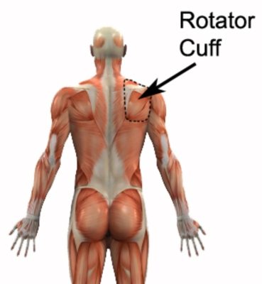
Supraspinatus anatomy plays a crucial role in maintaining optimal shoulder function and preventing injuries. It is one of the four rotator cuff muscles and the most commonly injured. By delving into the intricate details of this shoulder muscle, we can gain valuable insights into its origin, insertion, action, function, nerve supply, blood supply, palpation techniques, clinical relevance, common injuries, and find useful resources for further exploration. Join us as we explore the fascinating world of supraspinatus anatomy and discover how it impacts our overall shoulder health.
Origin and Insertion
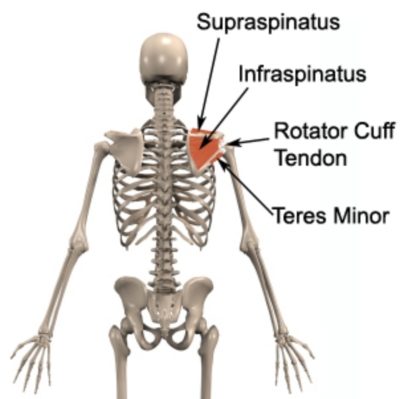
The supraspinatus muscle originates from the supraspinous fossa, a shallow depression located on the posterior and superior aspect of the scapula. From there, it forms a tendon that glides beneath the acromion, a bony projection of the scapula. The supraspinatus tendon attaches firmly to the greater tubercle of the humerus, a prominent bony prominence on the upper arm.
Action and Function
As a key player in the shoulder complex, the supraspinatus muscle primarily acts as an abductor of the shoulder joint. It initiates the initial 15 degrees of shoulder abduction, the sideways lifting of the arm from a relaxed position. Additionally, the supraspinatus muscle contributes to stabilizing the humeral head within the glenoid cavity during shoulder movement, serving as a safeguard against impingement and dislocation.
Nerve Supply
The supraspinatus muscle relies on the suprascapular nerve for its nerve supply. Emerging from the upper trunk of the brachial plexus, the suprascapular nerve traverses the suprascapular notch, providing innervation to both the supraspinatus and infraspinatus muscles.
Blood Supply
Optimal function and healing of the supraspinatus muscle are made possible by its arterial blood supply. The suprascapular artery, branching off from the subclavian artery, ensures the muscle receives the necessary oxygen and nutrients.
Palpation of Supraspinatus Anatomy
Palpation serves as a valuable tool for assessing the condition of the supraspinatus muscle and identifying potential abnormalities. By placing your fingertips just above the spine of the scapular (i.e. the supraspinous fossa) while the arm is relaxed, you can locate the supraspinatus muscle belly. Gentle pressure and palpation along the muscle’s course can help identify areas of tenderness, swelling, or muscle knots. The Supraspinatus insertion can be palpated by locating the tip of the acromion and palpating just below it – the greater tubercle of the humerus.
Supraspinatus Anatomy – Clinical Relevance
Understanding supraspinatus anatomy is vital in clinical practice. Various conditions can affect the supraspinatus muscle, leading to pain, weakness, and functional limitations. Common injuries involving the supraspinatus include tendonitis, partial or full-thickness tears, and impingement syndrome. These injuries often result from overuse, trauma, or degenerative changes and may require physiotherapy, rehabilitation exercises, or, in severe cases, surgical intervention.
Useful Links
For more in-depth information on related topics, you can explore the following articles on PhysioAdvisor:
- Rotator Cuff Tendonitis: Physiotherapy rehabilitation.
- Treating Rotator Cuff Tears: Physiotherapy and Surgical Interventions
- Shoulder Impingement Syndrome: Causes, Symptoms, and Treatment Options
- Shoulder Stretches and Shoulder Strengthening frequently used in rehabilitation
- Common Causes of Shoulder Pain and How to Manage Them
By deepening our knowledge of supraspinatus anatomy, we empower ourselves to better understand shoulder mechanics, prevent injuries, and facilitate effective treatment. Explore the links provided for a comprehensive understanding of related topics.
Supraspinatus Anatomy – References
- Drake RL, Vogl AW, Mitchell AW. Gray’s Anatomy for Students. 4th edition. Elsevier Health Sciences, 2019.
- Supraspinatus – Origin, insertion, innervation, action – by KenHub

Link to this Page
If you would like to link to this article on your website, simply copy the code below and add it to your page:
<a href="https://physioadvisor.com.au/supraspinatus-anatomy-origin-insertion-action-nerve-supply”>Supraspinatus Anatomy – PhysioAdvisor.com</a><br/>Learn about supraspinatus anatomy including origin, insertion, action, function, nerve supply, blood supply, injuries, palpation and more...
Return to the top of Supraspinatus Anatomy.