Tarsal Anatomy
Updated:
The tarsal bones make up the rear and midfoot. They play a crucial role in weight-bearing and movement of the foot and ankle. In this article, we will explore the anatomy of the tarsal bones, including their bony landmarks, muscular attachments, and common injuries.
Tarsal Anatomy: Bony Landmarks
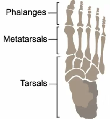
The tarsal bones consist of seven bones, which are:
- Calcaneus
- Talus
- Navicular
- Cuboid
- Cuneiforms (medial, intermediate, and lateral)
The calcaneus is the largest and strongest of the tarsal bones, forming the heel of the foot. The talus is the second-largest bone and is located between the calcaneus and the tibia (shin bone). The navicular bone is situated in front of the talus and is located on the medial (inner) side of the foot. The cuboid bone is located on the lateral (outer) side of the foot and forms the postero-lateral (rear / outer) aspect of the foot in front of the ankle. The cuneiform bones are located between the navicular bone and the metatarsals (bones in the midfoot) (Figure 1).
Tarsal Anatomy – Muscular Attachments:
Several muscles attach to the tarsal bones, including:
- Tibialis anterior: attaches to the navicular and cuneiform bones.
- Tibialis posterior: attaches to the navicular, cuneiform, and cuboid bones.
- Peroneus longus: attaches to the medial cuneiform and the base of the first metatarsal.
- Peroneus brevis: although this muscles doesn’t attach to the tarsal bones, it is held against the lateral surface of the calcaneus by the peroneal retinaculum and inserts into the 5th metatarsal.
Common Injuries affecting the Tarsal Bones:
The tarsal bones are susceptible to various injuries, including:
- Ankle Sprains: Ankle sprains occur when the ligaments that support the ankle are stretched or torn. These ligaments connect the tibia and fibula to the talus and calcaneus.
- Stress Fractures: Stress fractures are small cracks in the bones that occur due to overuse or repetitive stress. The navicular bone is a common site for stress fractures.
- Tarsal Tunnel Syndrome: Tarsal tunnel syndrome is a condition that occurs when the tibial nerve is compressed as it passes through the tarsal tunnel. This can cause pain, numbness, and tingling in the foot.
In conclusion, understanding the anatomy of the tarsal bones is important for anyone seeking to prevent or manage foot injuries. By knowing the bony landmarks, muscular attachments, and common injuries associated with the tarsal bones, individuals can take steps to protect their feet and maintain their overall health and well-being.
Tarsal Anatomy – References:
- Bones of the Foot – Tarsals, Metatarsals and Phalanges by Teach Me Anatomy: https://teachmeanatomy.info/lower-limb/bones/bones-of-the-foot-tarsals-metatarsals-and-phalanges/
- Saladin, K. S. (2011). Anatomy & Physiology: The Unity of Form and Function (6th ed.). New York, NY: McGraw-Hill.
- Magee, D. J. (2014). Orthopedic Physical Assessment (6th ed.). St. Louis, MO: Saunders.

Link to this Page
If you would like to link to this article on your website, simply copy the code below and add it to your page:
<a href="https://physioadvisor.com.au/tarsal-anatomy”>Tarsal Anatomy – PhysioAdvisor.com</a><br/>
Return to the top of Tarsal Anatomy.