Teres Minor Anatomy: Understanding Structure and Function
Updated:
The teres minor is a vital muscle located at the outer border of the shoulder blade and plays a crucial role in the mobility and stability of the shoulder joint. As we delve into the details of teres minor anatomy, we will explore its origin, insertion, action, nerve supply, blood supply, palpation techniques, clinical relevance, common injuries, and offer a list of useful links to relevant PhysioAdvisor articles for a comprehensive understanding.
Teres Minor Anatomy
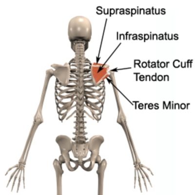
- Origin: The teres minor originates from the lateral border of the scapula, specifically from the upper two-thirds of the lateral border.
- Insertion: This muscle inserts into the greater tubercle of the humerus, blending with the posterior fibers of the deltoid muscle.
- Action: The primary function of the teres minor is to externally rotate the arm. Additionally, it aids in the stabilization of the humeral head within the glenoid cavity during shoulder movements.
- Function: Working in conjunction with other rotator cuff muscles, the teres minor helps in maintaining the overall stability and control of the shoulder joint during various activities involving arm movement.
Teres Minor – Nerve Supply and Blood Supply:
The teres minor muscle receives its nerve supply from the axillary nerve (C5-C6) and is part of the posterior cord of the brachial plexus. Its blood supply arises mainly from branches of the posterior circumflex humeral artery.
Teres Minor Anatomy – Palpation:
Palpating the teres minor can be a valuable technique for clinicians to assess its integrity and identify potential issues. To palpate the teres minor, the patient should be in a seated or lying position, with the arm slightly abducted ideally in a neutral position. The muscle can be located along the lateral (outer) border of the scapula, and its tendon can be felt as it attaches to the greater tubercle of the humerus.
Clinical Relevance:
- Common Injuries: The teres minor, being part of the rotator cuff, is susceptible to injuries, especially in sports that involve repetitive overhead movements like swimming, tennis, or baseball. The teres minor may also be injured due to repetitive or prolonged use of the shoulder and arm during everyday activities such as housework, lifting / carrying, pushing / pulling etc. Common injuries include strains, tears, and overuse syndromes, which can lead to pain, weakness, and limited range of motion.
- Rotator Cuff Tears: The teres minor and other rotator cuff muscles can be torn within their tendon or muscle belly.
- Rotator Cuff Impingement: Teres minor, along with other rotator cuff muscles, can get impinged between the acromion and humeral head, causing pain and discomfort.
- Tendonitis: Inflammation of the teres minor tendon can occur due to overuse or trauma.
Useful Links to physioadvisor.com.au Articles:
- Rotator Cuff Tear: Managing Shoulder Pain
- Rotator Cuff Tendonitis
- Shoulder Impingement: Causes and Treatment Options
- Rotator Cuff strengthening
Teres Minor Anatomy – Conclusion
In conclusion, understanding the anatomy of the teres minor is essential for healthcare professionals and individuals interested in shoulder health. Its role in shoulder stability and arm movement highlights its significance in maintaining upper limb functionality. While injuries to the teres minor can be challenging, proper diagnosis, treatment, and rehabilitation can help individuals recover and resume normal activities. Regular exercise, appropriate warm-up routines, and proper technique during physical activities can also prevent potential teres minor injuries, promoting overall shoulder health and well-being.
Teres Minor Anatomy – References:
- Teres Minor – Origin, insertion, action , innervation – by KenHub
- Moore KL, Dalley AF, Agur AMR. Clinically Oriented Anatomy. 7th ed. Philadelphia: Lippincott Williams & Wilkins; 2013.
- Drake RL, Vogl W, Mitchell AWM. Gray’s Anatomy for Students. 4th ed. Philadelphia: Churchill Livingstone; 2019.
- Standring S. Gray’s Anatomy: The Anatomical Basis of Clinical Practice. 41st ed. Philadelphia: Elsevier; 2016.

Link to this Page
If you would like to link to this article on your website, simply copy the code below and add it to your page:
<a href="https://physioadvisor.com.au/teres-minor-anatomy”>Teres Minor Anatomy: Understanding Structure and Function – PhysioAdvisor.com</a><br/>Learn about teres minor anatomy including origin, insertion, action, function, nerve supply blood supply, clinical relevance and more...
Return to the top of Teres Minor Anatomy: Understanding Structure and Function.