Tibia Anatomy: Understanding the Shin Bone
Updated:
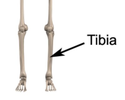
The tibia, also known as the shin bone, is a long bone located in the lower leg (figure 1). It is the second largest bone in the human body and plays an important role in weight-bearing and movement. In this article, we will explore the anatomy of the tibia, including its bony landmarks, muscular attachments, common injuries, and treatments.
Tibia Anatomy – Bony Landmarks
The tibia is a long bone that has several bony landmarks. The proximal end of the tibia is the largest part and has a triangular shape. The medial and lateral condyles are bony prominences on the top of the tibia that articulate with the femur bone to form the knee joint.
The tibial plateau is the flat surface on the top of the tibia where the femur rests. The tibial tuberosity is a bony prominence on the front of the tibia, just below the knee joint, where the patellar ligament attaches. The distal end of the tibia is also broad and forms the medial malleolus, which is the bony prominence on the inner aspect of the ankle joint.
Tibia Anatomy – Muscular Attachments
Several muscles attach to the tibia, including the quadriceps muscle, which attaches to the tibial tuberosity via the patellar tendon. The hamstring muscles also attach to the tibia, as well as the soleus and gastrocnemius muscles in the calf. These muscles are responsible for knee and ankle movement and stability.
The tibialis anterior muscle is another muscle that attaches to the tibia. It is located on the front of the shin and is responsible for dorsiflexion, or lifting the foot upwards.
Common Injuries affecting the Tibia
The tibia is susceptible to a variety of injuries, including fractures, stress fractures, and shin splints. Tibial fractures can occur due to trauma, such as a fall or car accident, or as a result of a sports injury. Stress fractures occur due to repetitive stress on the bone, such as in runners or other athletes.
Shin splints, also known as medial tibial stress syndrome, is a common injury that occurs due to overuse or improper training. It causes pain and inflammation along the inner edge of the tibia.
Treatment for tibia injuries may initially include rest, ice, compression, and elevation (RICE), physiotherapy, and in some cases, surgery. Physiotherapy can be particularly helpful for restoring leg strength, mobility and optimizing rehabilitation after an injury or surgery.
In conclusion, the tibia is an important bone in the lower leg that plays a crucial role in weight-bearing and movement. By understanding the bony landmarks, muscular attachments, and common injuries associated with the tibia, patients can better care for their legs and address any injuries that may arise. If you have any concerns about your leg health, be sure to consult with a qualified Physiotherapist.
Tibia Anatomy – References
- The Tibia by Teach Me Anatomy – The Tibia – Proximal – Shaft – Distal – TeachMeAnatomy
- Moore KL, Dalley AF. Clinically Oriented Anatomy. 7th ed. Philadelphia, PA: Lippincott Williams & Wilkins; 2014.
- Drake RL, Vogl AW, Mitchell AW. Gray’s Anatomy for Students. 2nd ed. Philadelphia, PA: Churchill Livingstone/Elsevier; 2010.
- Magee DJ. Orthopedic Physical Assessment. 6th ed. St. Louis, MO: Elsevier Saunders; 2014.

Link to this Page
If you would like to link to this article on your website, simply copy the code below and add it to your page:
<a href="https://physioadvisor.com.au/tibia-anatomy-understanding-the-shin-bone”>Tibia Anatomy: Understanding the Shin Bone – PhysioAdvisor.com</a><br/>Learn about tibia anatomy including bony landmarks, muscular attachments, common injuries and more on PhysioAdvisor
Return to the top of Tibia Anatomy: Understanding the Shin Bone.